
What is cortisol?
Cortisol is a steroid hormone that regulates a wide range of processes throughout the body, including metabolism and the immune response. It also has a very important role in helping the body respond to stress.
Cortisol is made in the cortex of the adrenal glands and then released into the blood, which transports it all round the body. Almost every cell contains receptors for cortisol and so cortisol can have lots of different actions depending on which sort of cells it is acting upon. These effects include controlling the body’s blood sugar levels and thus regulating metabolism, acting as an anti-inflammatory, influencing memory formation, controlling salt and water balance, influencing blood pressure and helping development of the fetus. In many species cortisol is also responsible for triggering the processes involved in giving birth.
Bound cortisol versus free cortisol:
80-90% of cortisol is bound to cortisol-binding globulin (CBG); much like thyroid is bound to thyroid-binding globulin (TBG) and testosterone is bound to sex hormone-binding globulin (SHBG).
A very small percentage of cortisol is free and unbound, while the remaining is in transition.
Converted and metabolized cortisol:
The human body produces cortisol first, and then different glands have the ability to keep it as cortisol or convert it into cortisone, which is biologically inactive, through the enzyme 11-beta-hydroxysteroiddehydrogenase (11bHSD).
Cortisol is then metabolized into 5-alpha-Tetrahydrocortisol (5a-THF) and 5-beta-Tetrahydrocortisol (5b-THF) and cortisone is metabolized into 5-beta-Tetrahydrocortisone (5b-THE).
Total metabolized output:
Since all production and output originally started as cortisol, the cortisone metabolites are added to the cortisol metabolites when evaluating the “total metabolized cortisol”. It essentially reflects how much cortisol was made in the body and has been processed out through the liver and the kidney into the urine.
Interpretation of different types:
The amount of cortisol produced and the amount of free cortisol available can be very different in some scenarios. Measuring both allows for insight into the rate of cortisol clearance/metabolism.
Higher levels of metabolized cortisol (vs free):
For example, higher levels of metabolized cortisol (compared to free cortisol) are often seen in obesity where adipose tissue is likely pulling cortisol from its binding protein and allowing for metabolism and clearance. The adrenal gland has to keep up with this cortisol sequestering and excretion, so cortisol production is often quite high (as seen in the levels of metabolized cortisol) even though free cortisol does not correlate positively with adipose tissue or BMI. This insight is quite helpful for those looking to lose belly fat and suspect cortisol/stress is a major factor. These people are often misdiagnosed as having low cortisol production when only free cortisol is measured. Increased cortisol clearance may also be seen in hyperthyroidism and is suspected to be part of the chronic fatigue story as well.
In people with low thyroid, the opposite pattern is often seen. When the thyroid slows down or if there is peripheral hypothyroidism where free T3 cannot get into the cells, the clearance (or metabolism) of cortisol through the liver slows down. As a result, free cortisol starts to increase and may show up elevated in urine.
The metabolized cortisol and free cortisol markers are important to use both together and separately in order to tell a more detailed story. Metabolized cortisol answers the question of how much cortisol is being made in total and clearing through the liver. Whereas free-cortisol results tell us how much cortisol is free to bind to receptors and allows for assessment of the circadian rhythm. The metabolites of cortisol also give insight into the relative activity of 11b-HSD types I and II, which controls the activation and inactivation (to cortisone) of cortisol.
How is cortisol controlled in general?
Blood levels of cortisol vary dramatically, but generally are high in the morning when we wake up, and then fall throughout the day. This is called a diurnal rhythm. In people that work at night, this pattern is reversed, so the timing of cortisol release is clearly linked to daily activity patterns. In addition, in response to stress, extra cortisol is released to help the body to respond appropriately.
The secretion of cortisol is mainly controlled by three inter-communicating regions of the body, the hypothalamus in the brain, the pituitary gland and the adrenal gland. This is called the hypothalamic–pituitary–adrenal axis. When cortisol levels in the blood are low, a group of cells in a region of the brain called the hypothalamus releases corticotrophin-releasing hormone, which causes the pituitary gland to secrete another hormone, adrenocorticotropic hormone, into the bloodstream. High levels of adrenocorticotropic hormone are detected in the adrenal glands and stimulate the secretion of cortisol, causing blood levels of cortisol to rise. As the cortisol levels rise, they start to block the release of corticotrophin-releasing hormone from the hypothalamus and adrenocorticotropic hormone from the pituitary. As a result the adrenocorticotropic hormone levels start to drop, which then leads to a drop in cortisol levels. This is called a negative feedback loop.
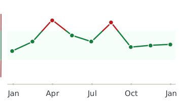
 a-Tetrahydrocortisol (a-THF) (male)
Every lab result you’ve ever had, charted and explained.
Healthmatters is a tracker for your lab results. Upload a report from any lab, any country — every marker charted and explained.
Start with my report →
All our content is backed by peer-reviewed studies, academic research, and trusted medical sources. We’re committed to accuracy and transparency — see our editorial policy for details.